the best probiotics
The small intestine and the immune system
The intestinal ecology is vital in both in the genesis, prevention and treatment of disease.
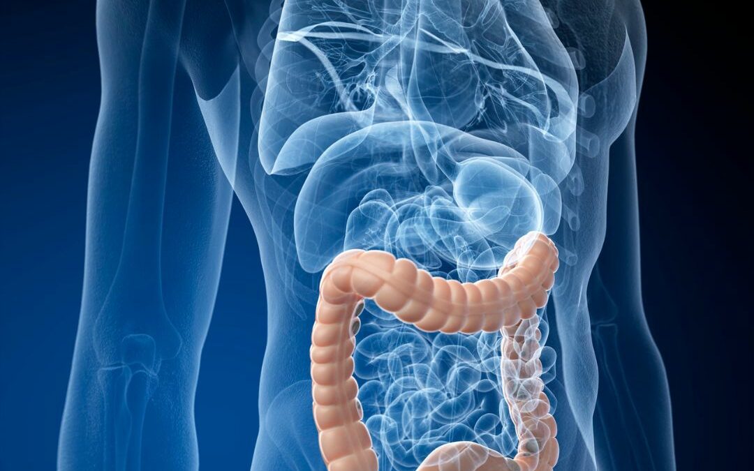
The mucosal membrane of the intestine, which has an area of around 200m2, is under constant challenge by different kinds of antigens. Not only do we swallow food antigens (60-70 tons throughout a lifetime) and food-borne microbes, but also inhaled particles that become trapped in the respiratory tract and transported up to pharynx are finally swallowed and reach the intestine. Furthermore, the mucosal membrane of the intestine has intimate contact with the normal intestinal microflora. It is therefore not surprising that the intestine contains the largest accumulation of lymphoid tissues in the body. Eighty percent (!) of the lymphocytes in the body are situated in the intestinal wall, in the so-called gut-associated-lymphoid-tissue (GALT). They appear as aggregates in lymph nodules called Peyer’s patches and as scattered lymphoid cells in the lamina propria. Further, the intestinal epithelia are seeded by small lymphocytes termed intraepithelial lymphocytes.
The GALT is associated with the rest of the lymphatic system via lymphatic vessels directed toward lymph nodules situated in the mesenteric fibrous tissue that lines the small intestine.
The immune system has two major functions, equally important: to react on foreign, dangerous antigens and particles like pathogenic bacteria and viruses and to not react on harmless antigens like food antigen and body tissue. These two functions have shown to be associated with each other.
Intestinal immune responses to bacteria are mainly induced in the Peyer’s patches. When lymphocytes (B- and T-cells) in the Peyer’s patches are activated by bacterial antigens they start proliferate and leave the intestinal tissue for the blood. They circulate during a couple of days before they settle down somewhere in the mucosa-associated-lymphoid-tissue (MALT), which is the lymphoid tissue associated with all the mucosal membranes in the body like the intestinal, respiratory and urogenital mucosa. Lymphoid tissues of all the mucosal membranes in the body are in this way interconnected. Immunological reactions, like allergic reactions in the intestine are likely to influence the status of other mucosal tissues. When the activated B-cells have settled down they mature and start producing IgA antibodies. Thus, the bacteria induce immunity against themselves. The paradox is that at the same time as the bacteria induce strong immunological reactions against themselves they decrease immunological reactivity against other antigens like food antigens. The mechanism of this is not completely known, but studies have shown that it is difficult to achieve oral tolerance in animals lacking an intestinal microflora while administration of bacterial antigens with the food increases the tolerating effect of feeding. Conversely some bacterial toxins like cholera toxin may break oral tolerance to food antigens so the composition of the intestinal flora is of importance.
Further, studies points to the importance of a higher turn-over of bacteria for the stimulation of the immune system. It is important that the immune system meet with new bacterial antigens continuously. As soon as IgA antibodies are produced against a certain bacterial antigen, antibodies bind to the antigen and inhibit it from translocating the intestinal wall and reach the Peyer’s patches. The low turn-over of bacteria and the decreased number of bacterial strains in the intestinal microflora of people in the western world may lead to a decreased bacterial stimulation of the immune system which could lead to over-reactivity against other antigens like food antigens.The immune system of an infant is not fully developed. The intestinal barrier is immature and the capacity to generate IgA-producing cells is inadequate. Bacterial colonisation provides maturational signals to the GALT. Thus, the intestinal microflora is important for the maturation of the immune system in infants. Animals lacking an intestinal microflora have very small Peyer’s patches, the level of IgA antibodies in blood is low and the amount of T-cells in the intestinal wall is also decreased.


